


по-русски
Modern spectroscopy methods in studying sctructure and function of biopolymers in biology and medicine
Русская версия |
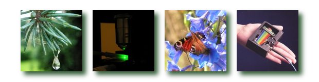
PresentationsOptical Biopsy of hard dental tissuesInstitute of electronics, Bulgarian Academy of Sciences,Tsarigradsko Shaussee 72, Sofia, 1784, Bulgaria, +359(2)9745742, E-mail: avramov@ie.bas.bg Sensitive methods that enable early detection and quantification of dental lesions increase possibilities to monitor changes in the tooth over time. Laser-induced fluorescence spectroscopy as a diagnostic method for detection of teeth lesions is used increasingly in recent years [1-2]. Easy and nondestructive fluorescence method for quantification of early caries improves clinical research and diagnostic possibilities. Fluorescence spectra of sound tooth and caries could be obtained when the tooth surface is illuminated with light in the blue - green spectral region. It is, therefore, very important to take into consideration not only the changes of the intensity, but also the changes of the shape of the fluorescence signal. Bearing in mind these variations, one will obtain a more complete picture of the pathologies investigated. The information contained in the spectral shape changes, related to the content of intrinsic fluorophores, allows a more accurate differentiation between carious and demineralized teeth. From the point of view of tissue optics, these results allow dental lesions to be considered as consisting of two different phenomena - tissue destruction and bacterial flora and its metabolism products increase. The results could be used to obtain a more complete picture of caries formation on the base of its fluorescent properties. On the basis of the results obtained in our investigations, the potential is demonstrated of the laser induced fluorescence method for non-invasive early diagnosis in dentistry [3]. These spectroscopic results could be applied in designing simplified fluorescence-imaging devices for detection and differentiation of the initial tooth caries from demineralized lesions.
|